The skeletal system includes all of the bones and joints in the human body. Every bone in the human body is a complex living organ that is made up of many cells, protein fibers and minerals. The skeleton acts like a scaffold by providing support and protection for the soft tissues that make up the rest of the body. The skeletal system provides connection points for muscels to allow movements at the joints. Brand new blood cells are produced by the red bone marrow inside of our bones.
Skull
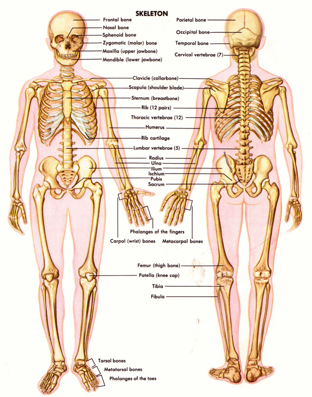
The skull is composed of 22 bones that are fused together except for the mandible. These 21 FUSED bones are seperate in kids to allow the skull and brain to grow, but fuse to give added strength and protection as an adult. The mandible remains as a moveable jaw bone and forms the only moveable joint in the skull along with the temporal bone.
Hyoid and auditory ossicels
The hyoid is a very small, U-shaped bone found just inferior to the mandible. The hyoid is the only bone in the body that does not form a joint with any other bone.
Vertebrea
Twenty-six vertebrea form the vertabral column of the human body. They are named by region:
Skull
The skull is composed of 22 bones that are fused together except for the mandible. These 21 FUSED bones are seperate in kids to allow the skull and brain to grow, but fuse to give added strength and protection as an adult. The mandible remains as a moveable jaw bone and forms the only moveable joint in the skull along with the temporal bone.
Hyoid and auditory ossicels
The hyoid is a very small, U-shaped bone found just inferior to the mandible. The hyoid is the only bone in the body that does not form a joint with any other bone.
Vertebrea
Twenty-six vertebrea form the vertabral column of the human body. They are named by region:
- cervical (neck) -7 vertabrea
- Thoriac (chest) - 12 vertebrea
- Lumbar (lower back) - 5 vertebrea
- sacrum - 1 vertebrea
- coccyx (tail bone) - 1 vertebrea
Ribs and sternum
The sternum, or breastbone, is a thin, knife-shaped bone located along the mid line of the anterior side of the thoriac region of the skeleton. The sternum connects to the ribs by thin bands of cartilage called the costal cartilage.
There are 12 pairs of ribs that together with the sternum form the ribcage of the thoracic region. The first seven ribs are known as “true ribs” because they connect the thoracic vertebrae directly to the sternum through their own band of costal cartilage. Ribs 8, 9, and 10 all connect to the sternum through cartilage that is connected to the cartilage of the seventh rib, so we consider these to be “false ribs.” Ribs 11 and 12 are also false ribs, but are also considered to be “floating ribs” because they do not have any cartilage attachment to the sternum at all.
Pelvic Girdle and Lower Limb
Formed by the left and right hip bones, the pelvic girdle connects the lower limb (leg) tot he axial skeleton.Formed by the left and right hip bones, the pelvic girdle connects the lower limb to the axial skeleton.The femur is the largest bone in the body and the only bone of the thigh (femoral) region. The femur forms the ball and socket hip joint with the hip bone and forms the knee joint with the tibia and patella. Commonly called the kneecap, the patella is special because it is one of the few bones that are not present at birth. The patella forms in early childhood to support the knee for walking and crawling.
The tibia and fibula are the bones of the lower leg. The tibia is much larger than the fibula and bears almost all of the body’s weight. The fibula is mainly a muscle attachment point and is used to help maintain balance. The tibia and fibula form the ankle joint with the talus, one of the seven tarsal bones in the foot.
The tarsals are a group of seven small bones that form the posterior end of the foot and heel. The tarsals form joints with the five long metatarsals of the foot. Then each of the metatarsals forms a joint with one of the set of phalanges in the toes. Each toe has three phalanges, except the big toe only has 2 phalanges.
pectoral girdle and upper limbs
The pectoral girdle connects the upper limb (arm) bones to the axial skeleton and consists of the left and right clavicles and left and right scapulae. The humerus is the bone of the upper arm. It forms the ball and socket joint of the shoulder with the scapula and forms the elbow joint with the lower arm bones. The radius and ulna are the two bones of the forearm. The ulna is on the medial side of the forearm and forms a hinge joint with the humerus at the elbow. The radius allows the forearm and hand to turn over at the wrist joint.
The lower arm bones form the wrist joint with the carpals, a group of eight small bones that give added flexibility to the wrist. The carpals are connected to the five metacarpals that form the bones of the hand and connect to each of the fingers.
Microscopic structure of bones
The skeleton makes up about 30-40% of an adult’s body mass. The skeleton’s mass is made up of nonliving bone matrix and many tiny bone cells. Roughly half of the bone matrix’s mass is water, while the other half is collagen protein and solid crystals of calcium carbonate and calcium phosphate. Living bone cells are found on the edges of bones and in small cavities inside of the bone matrix. Although these cells make up very little of the total bone mass, they have several very important roles in the functions of the skeletal system. The bone cells allow bones to:
Types of bones
Long-Long bones are longer than they are wide and are the major bones of the limbs. Long bones grow more than the other classes of bone throughout childhood and so are responsible for the bulk of our height as adults.
Short-Short bones are about as long as they are wide and are often cubed or round in shape. The carpal bones of the wrist and the tarsal bones of the foot are examples of short bones.
Flat-Flat bones vary greatly in size and shape, but have the common feature of being very thin in one direction. Because they are thin, flat bones do not have a medullary cavity like the long bones. The frontal, parietal, and optical bones of the cranium—along with the ribs and hip bones—are all examples of flat bones.
Irregular-Irregular bones have a shape that does not fit the pattern of the long, short, or flat bones. The vertebrae, sacrum, and coccyx of the spine—as well as the sphenoid, ethmoid, and zygomatic bones of the skull—are all irregular bones.
sesamoid- The sesamoid bones are formed after birth inside of tendons that run across joints. Sesamoid bones grow to protect the tendon from stresses and strains at the joint and can help to give a mechanical advantage to muscles pulling on the tendon.
Parts of bones
The long bones of the body contain many distinct regions due to the way in which they develop. At birth, each long bone is made of three individual bones separated by hyaline cartilage. Each end bone is called an epiphysis (epi = on; physis = to grow) while the middle bone is called a diaphysis (dia = passing through). The epiphyses and diaphysis grow towards one another and soon fuse into one whole bone. The region of growth and eventual fusion in between the epiphysis and diaphysis is called the metaphysis (meta = after). Once the long bone parts have fused together, the only hyaline cartilage left in the bone is found as articular cartilage on the ends of the bone that start form joints with other bones. The articular cartilage acts as a shock absorber and gliding surface between the bones to facilitate movement at the joint.
3 interesting facts about the skeletal system:
1.The smallest bone in our body is in your ear and its the size of a rice grain
2. Out of 206 bones in our body, 52 of them make both our feet up. (left, & right)
3. The longest bone in the body is the femur
The pectoral girdle connects the upper limb (arm) bones to the axial skeleton and consists of the left and right clavicles and left and right scapulae. The humerus is the bone of the upper arm. It forms the ball and socket joint of the shoulder with the scapula and forms the elbow joint with the lower arm bones. The radius and ulna are the two bones of the forearm. The ulna is on the medial side of the forearm and forms a hinge joint with the humerus at the elbow. The radius allows the forearm and hand to turn over at the wrist joint.
The lower arm bones form the wrist joint with the carpals, a group of eight small bones that give added flexibility to the wrist. The carpals are connected to the five metacarpals that form the bones of the hand and connect to each of the fingers.
Microscopic structure of bones
The skeleton makes up about 30-40% of an adult’s body mass. The skeleton’s mass is made up of nonliving bone matrix and many tiny bone cells. Roughly half of the bone matrix’s mass is water, while the other half is collagen protein and solid crystals of calcium carbonate and calcium phosphate. Living bone cells are found on the edges of bones and in small cavities inside of the bone matrix. Although these cells make up very little of the total bone mass, they have several very important roles in the functions of the skeletal system. The bone cells allow bones to:
- Grow and develop
- Be repaired following an injury or daily wear
Types of bones
Long-Long bones are longer than they are wide and are the major bones of the limbs. Long bones grow more than the other classes of bone throughout childhood and so are responsible for the bulk of our height as adults.
Short-Short bones are about as long as they are wide and are often cubed or round in shape. The carpal bones of the wrist and the tarsal bones of the foot are examples of short bones.
Flat-Flat bones vary greatly in size and shape, but have the common feature of being very thin in one direction. Because they are thin, flat bones do not have a medullary cavity like the long bones. The frontal, parietal, and optical bones of the cranium—along with the ribs and hip bones—are all examples of flat bones.
Irregular-Irregular bones have a shape that does not fit the pattern of the long, short, or flat bones. The vertebrae, sacrum, and coccyx of the spine—as well as the sphenoid, ethmoid, and zygomatic bones of the skull—are all irregular bones.
sesamoid- The sesamoid bones are formed after birth inside of tendons that run across joints. Sesamoid bones grow to protect the tendon from stresses and strains at the joint and can help to give a mechanical advantage to muscles pulling on the tendon.
Parts of bones
The long bones of the body contain many distinct regions due to the way in which they develop. At birth, each long bone is made of three individual bones separated by hyaline cartilage. Each end bone is called an epiphysis (epi = on; physis = to grow) while the middle bone is called a diaphysis (dia = passing through). The epiphyses and diaphysis grow towards one another and soon fuse into one whole bone. The region of growth and eventual fusion in between the epiphysis and diaphysis is called the metaphysis (meta = after). Once the long bone parts have fused together, the only hyaline cartilage left in the bone is found as articular cartilage on the ends of the bone that start form joints with other bones. The articular cartilage acts as a shock absorber and gliding surface between the bones to facilitate movement at the joint.
3 interesting facts about the skeletal system:
1.The smallest bone in our body is in your ear and its the size of a rice grain
2. Out of 206 bones in our body, 52 of them make both our feet up. (left, & right)
3. The longest bone in the body is the femur
